

Facial nerve paralysis is a condition characterized by partial or complete loss of movement of the muscles on one side of the face due to dysfunction of the facial nerve (cranial nerve VII). This nerve controls facial expression, eyelid closure, lip movement, and contributes to tear production, salivation, and taste sensation. Facial nerve paralysis can develop suddenly or gradually and may be temporary or permanent depending on the underlying cause and severity of nerve injury. Accurate diagnosis is essential to determine prognosis and guide appropriate management.
Causes
Facial nerve paralysis can result from a wide range of conditions affecting the nerve along its course from the brainstem to the facial muscles.
- Idiopathic causes: Bell’s palsy is the most common cause and is believed to be related to viral inflammation of the facial nerve
- Infections: Viral infections such as herpes simplex virus, varicella-zoster virus (Ramsay Hunt syndrome), Lyme disease, or otitis media
- Trauma: Temporal bone fractures, surgical injury, or penetrating facial trauma
- Tumors: Benign or malignant tumors involving the parotid gland, temporal bone, skull base, or cerebellopontine angle
- Neurologic conditions: Stroke, multiple sclerosis, or other central nervous system disorders
- Congenital causes: Developmental absence or malformation of the facial nerve
- Inflammatory or autoimmune disorders: Sarcoidosis or Guillain-Barré syndrome
The location and cause of nerve involvement significantly influence recovery potential.

Symptoms
Symptoms depend on the degree of nerve dysfunction and whether the paralysis is partial or complete. Onset may be sudden or progressive.
- Facial drooping: Inability to move one side of the face
- Inability to close the eye: Leading to dryness, irritation, or corneal injury
- Drooping of the mouth: Causing difficulty with speech and eating
- Loss of facial expression: Reduced ability to smile, frown, or raise the eyebrow
- Altered taste sensation: Especially involving the front portion of the tongue
- Excessive tearing or dry eye: Due to impaired eyelid function or tear production
- Facial pain or numbness: In select cases, depending on etiology
Chronic paralysis may lead to facial muscle tightness, asymmetry, or involuntary movements known as synkinesis.
Diagnosis
Evaluation focuses on determining the cause, location, and severity of nerve dysfunction. A detailed assessment is critical, particularly when paralysis is progressive or incomplete.
- Comprehensive head and neck examination: Including neurologic and otologic evaluation
- Facial nerve grading: Clinical scales may be used to assess severity and monitor recovery
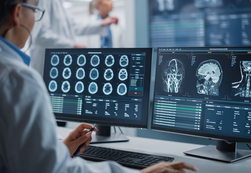
- Imaging studies: Magnetic resonance imaging (MRI) or computed tomography (CT) to evaluate the facial nerve, temporal bone, parotid gland, and brain
- Electrodiagnostic testing: Electroneurography or electromyography to assess nerve integrity and predict recovery
- Laboratory testing: Performed when infection, autoimmune disease, or systemic illness is suspected
Prompt evaluation is especially important when paralysis does not improve or worsens over time.
Treatment Options
Management depends on the underlying cause, timing of presentation, and degree of nerve injury. Early intervention can significantly influence outcomes.
- Medical therapy: Corticosteroids are commonly used in acute inflammatory causes; antivirals may be added in select cases
- Eye protection: Lubricating drops, ointments, eyelid taping, or temporary eyelid weights to prevent corneal damage
- Physical therapy: Facial muscle exercises and neuromuscular retraining to improve symmetry and control
- Surgical decompression: Considered in severe cases with evidence of significant nerve compression
- Facial nerve repair or grafting: Used when the nerve is transected or severely damaged
- Facial reanimation procedures: Static or dynamic surgical techniques to restore facial symmetry, movement, and function in long-standing paralysis
Treatment plans are individualized and may involve staged interventions over time.
When to See a Specialist
Evaluation by a head and neck or facial nerve specialist is recommended for any new facial weakness, particularly if onset is sudden, progressive, or associated with pain, hearing changes, or other neurologic symptoms. Early assessment improves diagnostic accuracy, protects eye health, and helps guide timely treatment. Patients with persistent or long-standing facial paralysis benefit from specialized care to address both functional limitations and quality-of-life concerns.